 Centers for Disease Control
and Prevention
11/20/2015 08:00 AM EDT
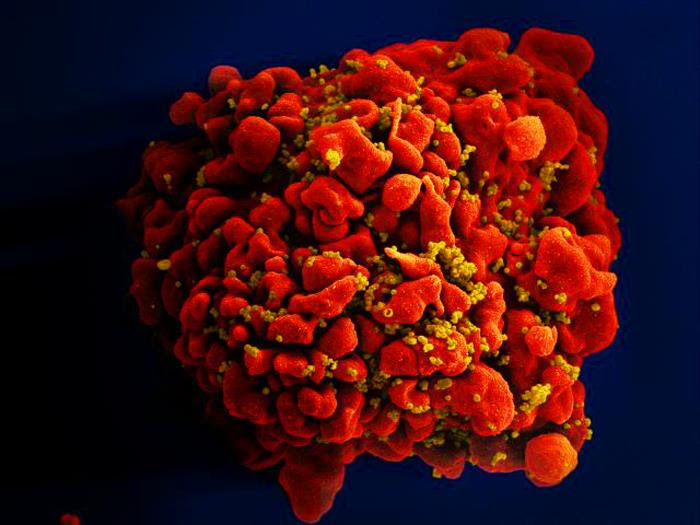
Produced by the National Institute of Allergy and Infectious Diseases (NIAID), this digitally-colorized scanning electron micrograph (SEM) depicts a single, red-colored H9-T cell that had been infected by numerous, spheroid-shaped, mustard-colored human immunodeficiency virus (HIV) particles, which can be seen attached to the cell's surface membrane.

11/19/2015 08:00 AM EDT
This 2007 photograph showed CDC biologist, Damian Danavall, preparing a "mastermix" for a multiplex real time PCR assay, which can differentiate between four sexually transmitted organisms known to be the cause for vaginal and urethral discharges.
11/18/2015 08:00 AM EDT
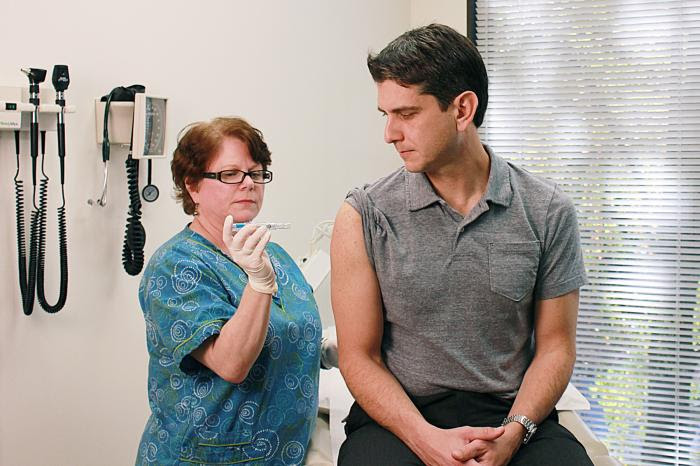
Taken inside a clinical setting, this qualified nurse had just administered a dosage of Fluzone intradermal influenza virus vaccine to a male clinic patient, using the patient's right shoulder region as the injection delivery site.

11/17/2015 08:00 AM EDT
This illustration depicts a three-dimensional (3D) computer-generated image of a number of rod-shaped, drug-resistant Shigella bacteria. The artistic recreation was based upon scanning electron micrographic imagery. Note that the exterior of the Shigella bacterium is fimbriated, covered by numerous thin, hair-like projections, imparting a furry appearance.

11/16/2015 08:00 AM EDT
This photograph was captured by U.S. Centers for Disease Control and Prevention (CDC) Health Communication Specialist, Alan Janssen, MSPH, while he was in the northern Indian state of Uttar Pradesh reviewing the methods, and implementation of the regions measles and polio vaccination programs.


No hay comentarios:
Publicar un comentario