Snapshots of Life: Green Eggs and Heart Valves
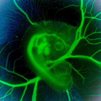
What might appear in this picture to be an exotic, green glow worm served up on a collard leaf actually comes from something we all know well: an egg. It’s a 3-day-old chicken embryo that’s been carefully removed from its shell, placed in a special nutrient-rich bath to keep it alive, and then photographed through a customized stereo microscope. In the middle of the image, just above the blood vessels branching upward, you can see the outline of a transparent, developing eye. Directly to the left is the embryonic heart, which at this early stage is just a looped tube not yet with valves or pumping chambers.
Developing chicks are one of the most user-friendly models for studying normal and abnormal heart development. Human and chick hearts have a lot in common structurally, with four chambers and four valves pumping two circulations of blood in parallel. Unlike mammalian embryos tucked away in the womb, researchers have free range to study the chick heart in or out of the egg as it develops from a simple looped tube to a four-chambered organ.
Jonathan Butcher and his NIH-supported research group at Cornell University, Ithaca, NY, snapped this photo, a winner in the Federation of American Societies for Experimental Biology’s 2015 BioArt competition, to monitor differences in blood flow through the developing chick heart. You can get a sense of these differences by the varying intensities of green fluorescence in the blood vessels. The Butcher lab is interested in understanding how the force of the blood flow triggers the switching on and off of genes responsible for making functional heart valves. Although the four valves aren’t yet visible in this image, they will soon elongate into flap-like structures that open and close to begin regulating the normal flow of blood through the heart.
When a valve forms incorrectly in a developing human, it can present a serious problem requiring major reparative surgery at a tender age. To discover ways to improve the early diagnosis and perhaps even prevention of these malformations, Butcher and his colleagues surgically induce heart defects to see in real time how it effects blood flow and subsequent development of heart valves. By combining their biomechanical studies with genomics, they’ve discovered that certain enzymes belonging to the GTPase family are activated in the chick by cyclic forces to coordinate the maturation of the mitral valve, which separates the heart’s left atrium and left ventricle chambers [1]. Without the timely activation of these enzymes, the mitral valve doesn’t develop properly.
These intriguing insights into early heart development hold promise for the roughly 40,000 babies born each year in the United States with congenital heart defects [2]. And that should give you food for thought the next time you reach for an egg. By the way, if you happen to be near NIH and want to see the 2015 BioArt winners in person, they are now on display free of charge at the Nobel Laureate Exhibit Hall inside the NIH Visitor Center.
References:
[1] Cyclic Mechanical Loading Is Essential for Rac1-Mediated Elongation and Remodeling of the Embryonic Mitral Valve. Gould RA, Yalcin HC, MacKay JL, Sauls K, Norris R, Kumar S, Butcher JT. Curr Biol. 2016 Jan 11;26(1):27-37.
[2] Congenital Heart Defects (Centers for Disease Control and Prevention)
Links:
Types of Congenital Heart Defects (National Heart, Lung, and Blood Institute/NIH)
Jonathan T. Butcher (Cornell University College of Engineering, Ithaca, NY)
Bioart (Federation of American Societies for Experimental Biology, Bethesda, MD)
NIH Support: National Heart, Lung, and Blood Institute


.png)


No hay comentarios:
Publicar un comentario