
Snapshots of Life: Fish Awash in Color
Credit: Chen-Hui Chen, Duke University
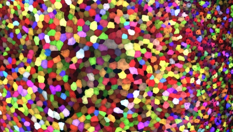
If this image makes you think of a modern art, you’re not alone. But what you’re actually seeing are hundreds of live cells from a tiny bit (0.0003348 square inches) of skin on the tail fin of a genetically engineered adult zebrafish. Zebrafish are normally found in tropical freshwater and are a favorite research model to study vertebrate development and tissue regeneration. The cells have been labeled with a cool, new fluorescent imaging tool called Skinbow. It uniquely color codes cells by getting them to express genes encoding red, green, and blue fluorescent proteins at levels that are randomly determined. The different ratios of these colorful proteins mix to give each cell a distinctive hue when imaged under a microscope. Here, you can see more than 70 detectable Skinbow colors that make individual cells as visually distinct from one another as jellybeans in a jar.
Skinbow is the creation of NIH-supported scientists Chen-Hui Chen and Kenneth Poss at Duke University, Durham, NC, with imaging computational help from collaborators Stefano Di Talia and Alberto Puliafito. As reported recently in the journal Developmental Cell [1], Skinbow’s distinctive spectrum of color occurs primarily in the outermost part of the skin in a layer of non-dividing epithelial cells. Using Skinbow, Poss and colleagues tracked these epithelial cells, individually and as a group, over their entire 2 to 3 week lifespans in the zebrafish. This gave them an unprecedented opportunity to track the cellular dynamics of wound healing or the regeneration of lost tissue over time. While Skinbow only works in zebrafish for now, in theory, it could be adapted to mice and maybe even humans to study skin and possibly other organs.
Skinbow was something of a serendipitous discovery. Several years ago, the Poss lab wanted to adapt a colorful neuroimaging technique in mice called Brainbow to study tissue regeneration in zebrafish. The lab’s interest was not to study neurons but to label cells permanently throughout the fish.
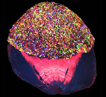
Caption: A scale from the Skinbow zebrafish showing the multicolored cells on its outer surface.
Credit: Chen-Hui Chen, Duke University
Credit: Chen-Hui Chen, Duke University
Chen inserted the Brainbow genes into several embryos to produce the desired research tool, but he soon noticed that one fish had developed a spectacular fluorescent pattern that was confined to the epithelial cells in the outer layer of the skin. Because the epithelial cells don’t divide, Chen and Poss saw a unique opportunity to study a large but relatively finite number of cells in an area of the body that was relatively easy to image. They also saw a chance to narrow their regenerative focus to the skin, which would allow them to image live cells and more readily visualize their movements and other transient behaviors involved in wound repair.
And so, Skinbow was launched. Each cell contains about 100 copies of the Brainbow gene cassette, and Poss estimates that cells could theoretically produce more than 5,000 distinguishably different colors. Interestingly, the genetically engineered fish have a reddish tint swimming in the aquarium when viewed with the naked eye. To see the Skinbow colors, the zebrafish are imaged with a regular confocal microscope under red, green, and blue light channels, and the images are then combined into works of art like the one above. If you want to see more of these cool images, take a look at this video.
References:
Multicolor Cell Barcoding Technology for Long-Term Surveillance of Epithelial Regeneration in Zebrafish. Chen CH, Puliafito A, Cox BD, Primo L, Fang Y, DiTalia S, Poss KD. Dev. Cell. 2016 Mar 21;36(6):668-680.
Links:
Learning About Human Biology From a Fish (National Institute of General Medical Sciences/NIH)
Zebrafish in the Classroom (University of Minnesota Duluth)
Tissue Engineering and Regenerative Medicine (National Institute of Biomedical Imaging and Bioengineering/NIH)
Kenneth D. Poss (Duke University School of Medicine/Durham, NC)
Brainbow (Center for Brain Science, Harvard University/Cambridge, MA)
NIH Support: National Institute of General Medical Sciences


.png)


No hay comentarios:
Publicar un comentario