12/20/2016 09:00 AM EST

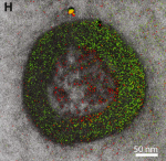
Seasons Greetings! What looks like a humble wreath actually represents an awe-inspiring gift to biomedical research: a new imaging technique that adds a dash of color to the formerly black-and-white world of electron microscopy (EM). Here the technique is used to visualize the uptake of cell-penetrating peptides (red) by the fluid-filled vesicles (green) of the […]
Merry Microscopy and a Happy New Technique!
 Seasons Greetings! What looks like a humble wreath actually represents an awe-inspiring gift to biomedical research: a new imaging technique that adds a dash of color to the formerly black-and-white world of electron microscopy (EM). Here the technique is used to visualize the uptake of cell-penetrating peptides (red) by the fluid-filled vesicles (green) of the endosome (gray), a cellular compartment involved in molecular transport. Without the use of color to draw sharp contrasts between the various structures, such details would not be readily visible.
This innovative technique has its origins in a wonderful holiday story. In December 2003, Roger Tsien, a world-renowned researcher at the University of California, San Diego (UCSD), decided to give himself a special present. With the lab phones still and email traffic slow for the holidays, Tsien decided to take advantage of the peace and quiet to spend two weeks alone at the research bench, pursuing an intriguing, yet seemingly wacky, idea. He wanted to find a way to deposit ions of a rare earth metal, called lanthanum, directly into cells as the vital first step in creating a new imaging technique designed to infuse EM with some much-needed color. After the holidays, when the lab returned to its usual hustle and bustle, Tsien handed off his project to Stephen Adams, a research scientist in his lab, thereby setting in motion a nearly 13-year quest to perfect the colorful new mode of EM.
Tsien, a co-recipient of the Nobel Prize in Chemistry in 2008, died unexpectedly last summer and is much missed. But before he died, Tsien received some fabulous news from Adams: their manuscript showing how well the new imaging method worked had been accepted for publication in the journal Cell Chemical Biology [1]. The technique, called multicolor electron microscopy, stands as yet another chapter in Tsien’s remarkable history of cell imaging advances, which includes his pioneering work in developing green fluorescent proteins as a research tool.
By bombarding molecules with electron beams, traditional electron microscopes can magnify the world inside of our cells up to 10 million times in black and white. With no optimal way to add color, researchers in the past have used heavy metals, such as lead and uranium, to scatter electrons in ways that add some contrast between the different molecular structures. But the level of resolution has remained less than ideal.
Now, multicolor electron microscopy offers a way use color to draw much clearer distinctions between molecular and cellular structures. For researchers who spend hours peering through microscopes, Adams notes, it’s like growing up watching black-and-white movies and one day walking into your first color matinee.
That takes us back to Tsien working in his lab over the winter holidays in 2003. Lanthanum is the prototype of a series of related elements called lanthanides. Tsien knew that if he could get lots of ionized lanthanides into cells, each metal in the series would lose its charge in a distinctive pattern that would make them easy to distinguish. If color coded and visualized on an electron micrograph, the metals could help to illuminate the inner workings of the cell.
As proof of principle in the Cell Chemical Biology paper, Tsien, Adams, and their UCSD collaborator Mark Ellisman succeeded in visualizing three lanthanides—lanthanum, cerium, and praseodymium—in distinctive red, green, and yellow. More colors are in the works, but, for now, here’s how the technique works:
Each ionized lanthanide is suspended in a clear solution that researchers paint sequentially onto the sample as it rests on the microscope. Although only two lanthanides at a time have been tried so far, the metals precipitate into the sample and, with the help of a linked molecule (diaminobenzidine), deposit in a place of research interest. The researchers then use a transmission electron microscope that distinguishes the locations of the metals by their unique energy-loss signatures. These data are then used to create maps of where the metals are in the sample, allowing the researchers to overlay this information as red and green on the usual black-and-white images of electron micrograph, as seen above,
Adams notes that Tsien spent just about every holiday in the lab, chasing his favorite ideas. His former lab members continue to work on some of them. So, there may be more to come to add to this Nobel Laureate’s rich legacy. Let’s hope so—and, in the meantime, Happy Holidays to you all!
Reference:
[1] Multicolor Electron Microscopy for Simultaneous Visualization of Multiple Molecular Species. Adams SR, Mackey MR, Ramachandra R, Palida Lemieux SF, Steinbach P, Bushong EA, Butko MT, Giepmans BN, Ellisman MH, Tsien RY. Cell Chem Biol. 2016 Oct 31. pii: S2451-9456(16)30357-30359.
Links:
Tsien Lab (University of California, San Diego)
Mark Ellisman (UCSD)
NIH Support: National Institute of General Medical Sciences; National Institute on Aging


.png)


No hay comentarios:
Publicar un comentario