06/15/2017 09:00 AM EDT

Last year, Nathan Krah sat down at his microscope to view a thin section of pre-cancerous pancreatic tissue from mice. Krah, an MD/PhD student in the NIH-supported lab of Charles Murtaugh at the University of Utah, Salt Lake City, had stained the tissue with three dyes, each labelling a different target of interest. As Krah […]

Snapshots of Life: A Van Gogh Moment for Pancreatic Cancer
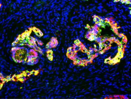
Last year, Nathan Krah sat down at his microscope to view a thin section of pre-cancerous pancreatic tissue from mice. Krah, an MD/PhD student in the NIH-supported lab of Charles Murtaugh at the University of Utah, Salt Lake City, had stained the tissue with three dyes, each labelling a different target of interest. As Krah leaned forward to look through the viewfinder, he fully expected to see the usual scattershot of color. Instead, he saw enchanting swirls reminiscent of the famous van Gogh painting, The Starry Night.
In this eye-catching image featured in the University of Utah’s 2016 Research as Art exhibition, red indicates a keratin protein found in the cytoskeleton of precancerous cells; green, a cell adhesion protein called E-cadherin; and yellow, areas where both proteins are present. Finally, blue marks the cell nuclei of the abundant immune cells and fibroblasts that have expanded and infiltrated the organ as a tumor is forming. Together, they paint a fascinating new portrait of pancreatic ductal adenocarcinoma (PDAC), the most common form of pancreatic cancer.
Pancreatic acinar cells, which produce and secrete digestive enzymes, are organized like a cluster of grapes, with a narrow stalk-like tube connected to them that is lined with duct cells. PDAC had long been described as arising in the duct cells. But recent studies show that acinar cells also can form PDAC tumors, inspiring several groups to try and figure out how that’s possible.
Krah and his colleagues in the Murtaugh lab think they might have the answer. It’s a protein called PTF1A, a transcription factor made only in acinar cells that turns genes on and off. Krah has found that PTF1A may be more than just a simple transcription factor. He and his colleagues propose that it is “a master regulator” that controls the activities of entire networks of genes, thereby giving acinar cells their unique and mature identity in the pancreas.
In studies of mouse and human cells grown in the lab, Krah and his colleagues have shown that acinar cells programmed to stop making PTF1A soon lose their cellular identities and convert into unstable ductal cells. When the loss of PTF1A is combined with the two most-common mutations in pancreatic cancer (KRAS and P53), the converted cells quickly turn pre-cancerous and, over time, will become full-blown cancer [1].
This work suggests that a previously undiscovered mechanism may be critical for suppressing the development of tumors in the pancreas. Because it can take 20 years or more for precancerous cells to develop into full-blown pancreatic cancer [2], this line of research may provide valuable leads on what to look for so this deadly type of cancer can be detected and treated much earlier. Now, that would be a truly beautiful and potentially life-saving outcome!
References:
[1] The acinar differentiation determinant PTF1A inhibits initiation of pancreatic ductal adenocarcinoma. Krah NM, De La O JP, Swift GH, Hoang CQ, Willet SG, Chen Pan F, Cash GM, Bronner MP, Wright CV, MacDonald RJ, Murtaugh LC. Elife. 2015 Jul 7;4.
[2] Distant metastasis occurs late during the genetic evolution of pancreatic cancer. Yachida S, Jones S, Bozic I, Antal T, Leary R, Fu B, Kamiyama M, Hruban RH, Eshleman JR, Nowak MA, Velculescu VE, Kinzler KW, Vogelstein B, Iacobuzio-Donahue CA. Nature. 2010 Oct 28;467(7319):1114-1117.
Links:
Pancreatic Cancer (National Cancer Institute/NIH)
Charles Murtaugh (University of Utah, Salt Lake City)
Research as Art (University of Utah)
NIH Support: National Cancer Institute


No hay comentarios:
Publicar un comentario