A Comparison of the Relative Detection and Quantification Techniques of Haemonchus contortus Eggs in Faeces Samples
Correctly identifying nematode eggs, especially when there are multiple species present, requires a high degree of expertise. As Strongylid eggs can look very similar, experts are required to differentiate the tiny differences in morphology, colour and egg shape that are used to identify different species.1
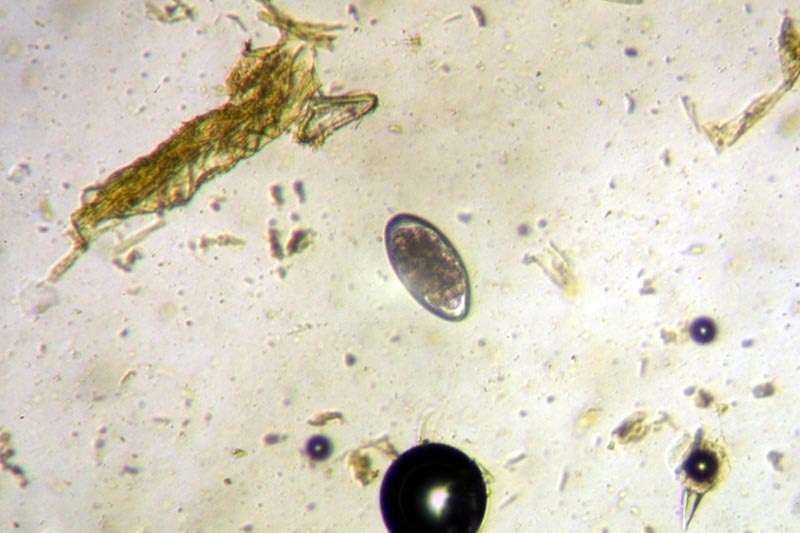
Credit: US Department of Agriculture
There are also issues with morphological examination in a standard diagnostic context as the eggs have to be viewed and quantified at a high magnification.2 Due to these drawbacks, lectin-binding assays with PNA (peanut agglutinin) have been developed and validated to specifically stain Haemonchus contortus eggs.3,4,5
However, fluorescent-labelled PNA has not been widely adopted by diagnostic labs because an advanced microscope equipped with fluorescent lights is required. This test is also time consuming, tedious to perform and PNA is difficult to quantify, which makes it difficult to justify as a diagnostic test when large quantities of samples need to be examined.
As animal trade increases between and within regions, correctly identifying H. contortus eggs in faecal samples has become increasingly important. In this study, to examine the available methods to detect and quantify H. contortus eggs, four identification methods were compared.
As a method of validating diagnostic test outcomes, the faeces of naturally infected flocks of 38 sheep were collected in Sweden. These were split into subsamples, which were analyzed with the following methods:
- Detection of DNA following amplification by real-time quantitative polymerase chain reaction (qPCR)
- Loop-mediated isothermal amplification (LAMP)
- Differential counting of eggs after staining with peanut agglutinin (PNA)
- McMaster egg counting
The MAST Group Ltd, designed and provided prototype pelleted LAMP reagents as an adaptation to the assay that Melville et al.6described. This assay was carried out “blind”.
The LAMP mastermix was produced using the protocol from the manufacturer; four pellets of reagent were resuspended in 344 μL 0.1 M Tris, 16 μL primer mix was added [1.6 μM FIP/ BIP, 0.8 μM FLP/BLP, and 0.2 μM F3/B3, these primer sequences are described by Melville et al.6] and 1 μL of template DNA was then added to every reaction, giving the final volume of 10 μL.
For the primer sequences, the LAMP reaction was kept in a real-time PCR machine (ABI 7500) incubated with the following conditions to maintain a consistent record of fluorescence and temperature over the total 33-minute reaction time: 60 cycles of 60°C for 1 second, and 61°C for 32 seconds. A FAM filter was used to record the fluorescence and the ABI 7500 software was used to analyze the results.
Spearman rank correlation, Bland-Altman plots and standard deviations were used to analyze the microscopic and molecular differences between similar tests. Strongylid egg counts had a range of 200-12,100 epg (eggs per gram; mean epg ± SD = 1,278 ± 2,049).
The presence of H. contortus eggs, were found to be present in 91% (34) in analysis with qPCR and 78% (29) tested positive in LAMP, while 76% (28) of PNA stained samples and 73% (27) of unstained samples detected the presence of eggs.
The Ct values of qPCR were between 25-49 (mean ± SD = 33 ± 6), while the LAMP Ct (cycle threshold) values were between 13-38 (mean ± SD = 21 ± 7). In the qPCR and LAMP analyses, three (8%) and seven (19%) samples, respectively, had a Ct of over 35, whereas no reactions were observed in three (8%) and eight (22%) samples, respectively.
Microscopic examination and DNA detection showed that the diagnostic tests showed a good agreement, with the molecular tests being more sensitive. The molecular tests showed a larger bias (−9.8 ± 10) than with the microscopy methods (−4.2 ± 11).
Test sensitivity was observed to be ranked thus: qPCR > LAMP > PNA > McMaster counting using conventional microscopy.
Overall it was found that McMaster counting could be employed to identify H. contortus without major mistakes such as results from false positives. However, increased accuracy to diagnose H. contortus was provided by the molecular methods.
This becomes essential during quarantine investigations or antihelmintic treatment efficacy studies and can be applied when studying faecal matter of wildlife to examine nematode transmission from the wild to livestock.
While the study did not originally set out to examine the practicalities behind the set up and commercialization of these techniques in a smaller diagnostic setting, LAMP seems a particularly viable option in even a smaller diagnostic lab, simply because it can provide reliable and highly sensitive results in under sixty minutes.
This is in contrast to qPCR, which has a higher detection rate, but takes longer than a LAMP assay to run. LAMP could easily be used as a validation technique rather than the more tedious and complicated egg-counting PNA.
References
- Georgi JR, McCulloch CE. Diagnostic morphometry: identification of helminth eggs by discriminant analysis of morphometric data. Proc Helm Soc Wash (1989) 56:44–57.
- Roeber F, Kahn L. The specific diagnosis of gastrointestinal nematode infections in livestock: larval culture technique, its limitations and alternative DNA-based approaches. Vet Parasitol (2014) 205:619–28. doi:10.1016/j. vetpar.2014.08.005
- Jurasek M, Bishop Stewart J, Storey B, Kaplan R, Kent M. Modification and further evaluation of a fluorescein-labeled peanut agglutinin test for identification of Haemonchus contortus eggs. Vet Parasitol (2010) 169:209–13. doi:10.1016/j.vetpar.2009.12.003
- Palmer DG, McCombe IL. Lectin staining of trichostrongylid nematode eggs of sheep: rapid identification of Haemonchus contortus eggs with peanut agglutinin. Int J Parasitol (1996) 26:447–50. doi:10.1016/0020-7519(96)00009-4
- Colditz IG, Le Jambre LF, Hosse R. Use of lectin binding characteristics to identify gastrointestinal parasite eggs in faeces. Vet Parasitol (2002) 105: 219–27. doi:10.1016/S0304-4017(02)00013-4
- Melville L, Kenyon F, Javed S, McElarney I, Demeler J, Skuce P. Development of a loop-mediated isothermal amplification (LAMP) assay for the sensitive detection of Haemonchus contortus eggs in ovine faecal samples. Vet Parasitol (2014) 206:308–12. doi:10.1016/j.vetpar.2014.10.022
About Mast Group
Mast is an independent world class manufacturer and supplier of diagnostic products for clinical, industrial and veterinary testing.
The parent company, Mast Group Ltd., has corporate headquarters in Bootle near Liverpool, UK. Mast also has Subsidiary companies in Reinfeld, Germany (Mast Diagnostica GmbH) and Amiens, France (Mast Diagnostic).
Manufacturing of the core range of microbiology products takes place in Bootle while other infectious disease and autoimmune diagnostics, using a variety of technologies, are produced in Reinfeld.
Mast offers its extensive product range through its own companies plus a global network of Distributors, for example, Mast has a long standing association with Davies Diagnostics in Johannesburg, South Africa.
Mast also supplies on a country by country basis, other quality diagnostic products from several internationally renowned manufacturers.
Sponsored Content Policy: News-Medical.net publishes articles and related content that may be derived from sources where we have existing commercial relationships, provided such content adds value to the core editorial ethos of News-Medical.Net which is to educate and inform site visitors interested in medical research, science, medical devices and treatments.
Last updated: Mar 6, 2018 at 9:51 AM


.png)


No hay comentarios:
Publicar un comentario