Applications of FISH
Fluorescent in situ hybridization (FISH) is a genetic technique used to diagnose congenital diseases such as Down's Syndrome and Edward's Syndrome. It has also been used to detect cancer and diagnose infectious diseases.

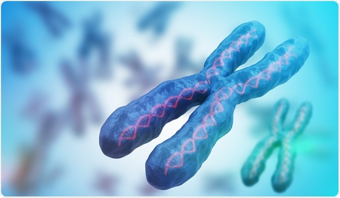
Credit: vchal/Shutterstock.com
The DNA in our cells consists of a double helix or two strands of DNA which are repeatedly wound round each other. Each strand contains a sequence of the four bases: adenine (A), guanine (G), cytosine (C), and thymine (T). Each base in a strand pairs to its complementary base on the other strand (A pairs with T, C pairs with G) using hydrogen bonding.
The high affinity with which of complementary base pairs (A-T, C-G) bind to their corresponding partner is called ‘hybridisation’. Hybridisation forms the basis of Fluorescence In Situ Hybridisation or FISH.
In FISH, small DNA strands which have been labelled with a fluorescent probe are allowed to bind to fragments of a person’s DNA, corresponding to a particular genomic region of interest. This is useful to assess chromosome or gene copy number.
If there has been a duplication, more of the probe will hybridise with the sample DNA and more fluorescence will be detected. If there has been a deletion in the genome, the probe cannot attach to its complementary region as it is absent and no fluorescence will be detected. This technique has been applied to serve diagnostic purpose in clinical medicine.
Prenatal diagnosis of chromosomal abnormalities
Prenatal diagnosis is critical to determine the health and congenital abnormalities in the unborn fetus. Common chromosomal abnormalities include Patau syndrome (presence of an extra copy of chromosome 13), Edward’s syndrome (presence of an extra copy of chromosome 13), and Down’s syndrome (presence of an extra copy of chromosome 21).
When a florescent DNA probe is tested against healthy regions of chromosome 13, 18, or 21, two fluorescent signals corresponding to two chromosomes would be observed. However, in the case of gene duplication or presence of extra copy of chromosome, three florescent signals would be visible. Thus, FISH has led to easy diagnosis of congenital disorders which may not easily evident under a microscope.
Detection of copy number variants (CNVs) in adults
Different types of FISH probes can detect different chromosomal aberrations. Probes specific to a particular gene locus can determine gene fusions, aneuploidy or abnormal number of chromosomes, and loss of chromosomal region or whole chromosome in an adult.
FISH has 50-fold higher resolution than the conventionally used Giemsa stain to detect genetic diseases. The Giemsa stain compares the light and dark patterns of euchromatin and heterochromatin between control and test case to detect defects; whereas FISH is based on direct visualization of specific regions of genome. Also, it is less time consuming than karyotyping which requires culturing of cells and subsequently arrest at metaphase to observe the structural features of chromosomes.
Detection of cancer
FISH is widely used to detect the chromosomal abnormalities associated with cancers. HER2 positive breast cancer has extra copies of HER2 gene and FISH testing is done on breast cancer tissue removed during biopsy to detect the extra copies of the gene. Additional copies of the HER2 gene lead to more HER2 receptors in the cells and these receptors receive signals that stimulate the growth of breast cancer cells.
FISH also has higher sensitivity in detecting cancers. In a case of bladder cancer, FISH has 50% higher sensitivity (true positive rate) than routine cytology. Chromosomal aberrations are found in 60% of acute myeloid leukemia and 65-90% of acute lymphocytic leukemia. FISH was found to more sensitive and reliable technique to diagnose acute leukemia compared to G-banding which involves staining the chromosomes with Giemsa stain to yield dark and light bands which correspond to heterochromatin and euchromatin.
Detection of infectious diseases
Phylogenetic group-specific 16S ribosomal RNA (rRNA) are used in FISH to detect infectious agents. Oligonucleotide sequences complementary to 16S rRNA (17-34 nucleotides in length) as FISH probes can be used for the analysis of microbial communities in oral cavity and gastrointestinal flora. Both oral cavity and intestine are heavily colonized, and FISH has been used to detect pathogens within the microbial community.
Specific oligonucleotide probes have also been designed to detect pathogens in respiratory tract infections. Similarly, FISH has also been used to detect pathogens within tissues. Genus and species specific oligonucleotide probes have been used to identify pathogenic microorganisms in blood cultures. For example, FISH probes complementary to specific sequence of 16s rRNA can detect malaria infection in blood samples.
Reviewed by Kate Anderton, BSc
Last Updated: Apr 3, 2018


.png)


No hay comentarios:
Publicar un comentario